Contacts. pesteducation.com, (714) 960 8022, weevilways@earthlink.net,
Flea Biology and Control
By Dr.
Richard and Patricia Kaae
Adult
fleas are small, wingless critters with backward projecting spines on their
flattened highly sclerotized (hard) body. All these characteristics are well
suited for their ectoparasitic way of life. Of course, there would be no
advantage in being large as the host could easily remove such a creature. Insect wings are generally most useful in
either finding a mate, food or seeking a favorable environment and all these
factors seem to be readily available on the flea’s host. The backwards-projecting spines make it more
difficult for the host to scratch-out or remove an adult flea because the
spines can readily lodge in hair or feathers. Of course, forward projecting
spines would hinder the flea’s movement through the forest of hair of many
hosts. The adult flea is flattened from side to side thus allowing it to easily
move through the host. Finally, a heavy
sclerotized or thick exoskeleton makes it more difficult for the host to kill
adult fleas by scratching or biting.
Fleas
have a limited sense of smell with small antennae concealed in grooves.
However, they can readily detect carbon dioxide thus possibly explaining why
fleas get so excited and jump wildly when coming in contact with breath. Depending on the species, fleas may or may
not have eyes but even in the case of the former they are thought to have a
poor sense of vision, which is limited to detecting changing pattern of light
and shadows. Their bodies are covered by
a variety of sensory hairs and spines that can detect the vibrations of other
animals. As a result of these various structures
fleas are well adapted for detecting a host using smell (CO2), vibrations and
shadows. This ability was recently
illustrated by an experiment conducted on rabbit fleas. Two hundred and seventy-five adult fleas were
marked and randomly released in an 18,000 square foot enclosed field. Then three rabbits were released in the
field. After a few days over half of the released fleas were found on the
rabbits.
FLEA IDENTIFICATION
Worldwide
there are over 1,600 species of fleas while in the US there are a few
hundred. Most species are somewhat host
specific and this alone can be used to a degree for identification (rabbit
fleas occur mainly on rabbits, mice fleas occur primarily on mice, etc.). Regardless of this large number some species
are much more commonly encountered and of more economic importance than
others. The following is meant to be a
beginning source for the identification of some of the more common economic
species. The main criteria that are used
for species identification in fleas are the presence or absence and shape of
the pronotal and genal combs (Figure 1), length of the labial palps, shape of
the head and a few other minor morphological characteristics.

Figure 1.
The head and thorax of a cat flea illustrating the teeth of the genal
and pronotal combs. Images courtesy of
Department of Parasitology, University of Sao Paulo, Brazil.
Based
on structural differences the fleas that are commonly encountered in the US
fall into 3 groups, mainly those which possess a genal and pronotal comb, those
that only have a pronotal comb and those that lack both types of combs. The
following key can be used to identify many of the common fleas found in the US.

Fleas with a Genal and Pronotal Comb
The
cat flea (Ctenocephalides felis) and the dog flea (Ctenocephalides
canis) obviously belong to the same genus and are very similar in
appearance. They can be distinguished
from other common fleas by the presence of both genal and pronotal combs. The mouse flea and rabbit fleas share this
characteristic and can be distinguished from the cat and dog flea later. The main characteristic that is used to
distinguish a cat flea from dog fleas is that in the dog flea the first tooth
on the genal comb is distinctly shorter than the second while in the cat fleas
they are of equal length. (Figure 2).

Cat Flea
Dog Flea
Figure 2.
A comparison of the cat and do fleas.
Note in the cat flea the first and second teeth on the genal combs are
of equal length, while in the dog flea the first is significantly shorter than
the second. Images courtesy of
Department of Parasitology, University of Sao Paulo, Brazil.
As
previously mentioned the rabbit and mouse fleas also possess a genal and pronotal
comb. The mouse flea can be distinguish
from the above 2 fleas and the rabbit flea by the fact that it lacks eyes
(Figure 3).

Figure 4. A mouse
flea (Leptopsylla segnis).
Distinguished by the presence of a genal and pronotal comb and absence
of an eye.
As
with the above 3 species, the rabbit flea possesses a genal and pronotal comb
but the distinguishing characteristic is that the genal comb is almost vertical
in the rabbit flea (Figure 54) as
opposed to a horizontal position in the other 3 species. The teeth on the comb are blunt in the rabbit
flea.
Figure 4. A
rabbit flea with blunt teeth on a nearly vertical genal comb.
Common Fleas with Only a Pronotal Comb
The
northern rat flea (Figure 5) and the ground squirrel flea possess only a
pronotal comb. No image is available for
the ground squirrel flea.

Figure 5. The
northern rat flea, Nosopsyllus fasciatus, is distinguished by lacking a
genal comb.
Common Fleas Lacking Both a Genal and Pronotal Comb.
The stick tight
flea (Figure 6) is characterized by the absence of a genal and pronotal comb
and presence of an angular head and contracted thoracic area.

Figure 6. Stick tight flea. Image courtesy of Department of Parasitology,
University of Sao Paulo, Brazil.
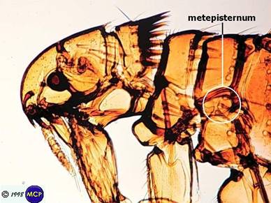
The Oriental
rat flea, Xenopsylla chelopis, (Figure 7) is characterized by the
absence of a genal and pronotal comb, and the mesonotum is divided by a
thickening and the ocular bristle inserted in front of the eyes.
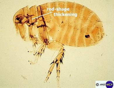
Figure 7.
Oriental rat flea. Image courtesy of
Department of Parasitology, University of Sao Paulo, Brazil.
The human
flea, Pulex irritant (Figure 8)
is characterized by a lack of genal and pronotal combs, and no
mesonutum division and the ocular bristle located below the eye.

Figure
8. Human flea. Image courtesy of
Department of Parasitology, University of Sao Paulo, Brazil.
FLEA BIOLOGY
Fleas are often found on birds and mammals with over 2,000-3,000 species occurring worldwide. Based on feeding habits, there are 3 main types of fleas. The first type does not attach to the host while feeding. These fleas are easily transmitted from one host to another and most belong in this category. The second type includes stationary females that are anchored by their mouthparts while feeding on the host. The third includes gravid females that develop under the host's skin and maintain a breathing pore to the outside.
The life cycles of most common
fleas found in the US are somewhat similar.
The following is based on the most common flea found on pets, namely the
cat flea, Ctenocephalides felis (Figure 2). As with most fleas in
temperate areas of the world cat fleas infestations tend to be seasonal
reaching peak population in the summer months and in the colder areas dropping
to almost non-existent in the winter months.
As with all insects the length of the life cycle and therefore rate of
reproduction is directly proportional to prevailing temperatures. To a point
the warmer the temperature the faster an insect completes its life cycle. Also outdoors colder temperatures and rain
greatly increase the mortality rate of fleas.
Of course if pets are confined to indoor conditions or someone live in
warmer tropical areas flea season can be year around.
The flea spends most of its
adult stage on the host. As with most
fleas the cat flea has preferred hosts (dog and cats) but in the absence of
these will find and feed on other warm-blooded animals, including humans.
The mouthparts of adult fleas are adapted for
slicing the skin and siphoning blood. In
order to produce eggs a female flea must have a daily blood meal. Once fed she
typically deposit her eggs on the host but most will drop off, as they are
relative dry. The eggs are relatively
large for such small insects, about the size of a grain of salt or
approximately 1/12th the size of the adult flee. They are oval and rounded on both ends. A female flea deposits several eggs a day but
since they are long lived she is capable of producing 300 to 400 eggs during
her lifetime. When an infested pet (such
as a cat during fleas season) sleeps on a table or other surface and leaves, it
almost looks as if someone has taken salt and pepper and sprinkled it on this
surface. This is called the salt and
pepper effect. Of course the salt is the
fleas’ eggs and the pepper is the feces of the adult fleas.
Once deposited the eggs hatch
in 2 to 12 days depending on prevailing temperatures. As previously discussed the warmer the
prevailing temperature (to a point) the faster the eggs hatch. Optimum development for fleas typically
occurs in locations protected from rainfall and sunlight with a relative
humidity of at least 75% and temperatures between 70 to 90 F.
The flea larva is elongated,
legless with a well-developed head capsule and elongated sparse hairs on the
body. When disturbed they will
characteristically flip in circles. The
larval stage (Figure 9) is a scavenger feeding on any of a variety organic
matter including dander, feces, food particles and other organic matter. One of main ingredients in their diet is the
adult feces that is quite high in protein.
Adult fleas bite and feed many more times than are needed to fulfill
their nutritional requirements. As a
result their feces is very high in partially or undigested blood. This is a very efficient system. As the host walks around it is not only
dropping flea eggs into the environment but is also dropping the main
ingredient of the larval diet.

Figure 9. The
larval stage of a flea, a scavenger.
Image courtesy of CDC Healthwise Photo Library.
The
larval stage may be completed in as little as 9 days or under unfavorable
conditions can be extended as long as 200.
Larval development is
normally restricted to protected places where there is at least 75% relative
humidity. At the end of this active feeding stage after the larva has
reached full growth, it spins a cocoon around its body and pupates. The pupal cocoon (Figure 10) typically is
covered by bits of their environment including such materials as lint, hair,
dirt and sand. Again the length
development of the pupal stage is quit variable but at room temperature
approaches 2 weeks.


Figure 10. The
pupa in case (left). Pupa (right) removed from case. Image courtesy of Department of Parasitology,
University of Sao Paulo, Brazil.
Once fleas emerge from the
pupae they remain in the silken cocoons until the presence of an approaching
host trigger them out. Prior to emergence from the cocoon an adult flea can
survive for an extended period of time (350 days or more). However, once emerged the adult must feed or
it will die in a few days. If
continuously fed some species can live for a year or more. The longest recorded
life cycle of a flea is 996 days for Pulex irritans, the human flea.
FREQUENTLY ASKED QUESTIONS
A few occurrences or questions
homeowners commonly encounter or ask can be explained by flea biology.
Scenario #1. Prior to taking their German Shepard with
them on a 4-week vacation, homeowners have a little problem with flea bites.
They return to find dozens of fleas jumping all over their legs. Answer.
Prior to leaving, most of the fleas that emerged from their pupae fed on
the preferred host--the dog. While they
were gone, many fleas completed their life cycle as the host presence was not
necessary; the larvae are scavengers.
Because no one was home, the adult fleas that emerged remained in their
loose cocoons. Upon their return, the
homeowners’ vibrations triggered the fleas out of their cocoons. Of course one solution would be to send the
dog in first to collect all the waiting fleas (joke).
Scenario #2. Someone has a flea
problem in their home but does not have any pets. In this case, there was probably a
flea-ridden animal in or under the home at one time. Upon its departure the
fleas were left to seek the only available host, the human. Possibly someone had spent a few days in the
home with their pet dog or possibly there was a litter of kittens below the
house. There may not be many fleas in
the home with this type of situation, but those that are left will make their
presence known and will bite the homeowner.
Question #1. I am frequently bitten by fleas and my wife
is almost never attacked. Why?
Answer. Blood feeding insects
such as fleas and mosquitoes are attracted to their victim by chemicals such as
those found in sweat and breath (CO2).
People obviously smell different and are more or less attractive. Amazingly, there even seems to be a
difference in attractiveness to individuals depending on the geographical
area. We have noticed that mosquitoes
are more attracted to my wife in Thailand but are more attracted to me in
Central America. Individual sensitivity
to bites is another factor, which can partially answer this dilemma. To some bites are extremely annoying while to
others they are hardly noticed. The
difference is due to an allergic reaction to the saliva that is injected when a
flea feeds. There are different degrees
of allergic reactions amongst different individuals. The function of the saliva is to keep blood
flowing while the flea feeds. It
contains an anticoagulant.
Question #2. I just moved into an apartment and there were
hundreds of fleas. This is especially
puzzling since it has been vacant for 2 months and it was treated for fleas when
the previous tenant moved. Answer. The people who lived there before you
probably had pets. The adult fleas you
are seeing are those that have not emerged from their cocoons. Remember adult fleas can remain alive in this
condition for up to 12 months or so. If
the house was previously treated, treatment probably did not penetrate the
carpet where these larvae and pupae were developing.
Question #3. I have bombed my house three times this month
and still have a problem with fleas.
Why? Insecticide bombs do not to
penetrate and would not reach developing flea larvae and pupae deep in the
rugs. If you did not treat your yard it
is possible that new adult fleas could be coming into your house on you or your
pet.
Questions
#4. Why do fleas seem to be more of a
problem in my beach house as opposed to my home in the inland valley? Answer.
First of all part of the answer may be due to differences in
climate. Temperatures at the beach tend
to be milder and therefore favorable for flea development and survival than
those in inland areas. Beach areas tend
to also have a higher humidity, which is also favorable for flea development
and survival. Finally people frequently
confuse sand fleas with true fleas.
Question #5. What are sand fleas? "Sand fleas"
or "beach fleas" are common names for small orange crustaceans called
amphipods found along the beach. They are distant non-insect relatives of true
fleas, and do not even bite. Also, some people may refer to fleas that just
happen to be developing in sandy areas as "sand fleas".
FLEA DISEASE AND OTHER MALADIES.
The main rat
during the major plagues of the world was the so called black rats (Rattus
rattus). Today this is better known as the roof rat. It was the
dominant rat during the now historical plagues.
Today the dominant rat in many areas of the world is the Norway rat (Rattus norvegicus). The black or roof
rat is more condusive to living indoors and was a better climber than the
heavier bodied Norway rat. In addition they rarely moves more
than 200 meters from their nest, hence their adaptability to the thatched roof
homes of either the Middle Ages, present day rural Africa, or parts of the
Asian subcontinent. Normally the fleas live on the rodents in a form of
equilibrium, but sometimes that equilibrium is upset when they multiplies rapidly in the flea's gut,
eventually blocking the lumen (the space within its gut) so that the flea
regurgitates infected material as it attempts to feed. This infects the rodent
and it contracts a form of the fatal disease called silvatic plague. When infected, rats are asymptomatic until near
death, whereupon they swell up (because the Y. pestis grow so rapidly
and in such large numbers) and stagger as if intoxicated. The fleas then leave
their dying hosts and seek residence in the nearest warm-blooded animals. In
the case of the major plagues of the world this was frequently a human.
There are 3 forms of plague, namely bubonic,
pneumonic, and septicemic. All are caused by the bacterium, Yersenia pestis. Each form kills individual in different
ways. With bubonic plague 1 to six days after a human receives a flea bite, the
lymph nodes in the armpit and groin become very tender and swollen ranging in
size from that of a egg to apple. These very painful swollen areas are called buboes. The buboes may break and discharge
pus.
Occasionallythe original bite site becomes infected
and may turn gangrenous and necrotic (rotting). Other symptoms are headaches,
nausea, aching joints, fever of 101-105 degrees, vomiting, and a general
feeling of illness. Symptoms take from 1-7 days to appear. Then if the fever
breaks, there is usually remission and the vicitms’s immune system takes over
eliminating the bacterium and symptoms. If the fever doesn't break, the
infection spreads to the blood, causing septicemia and death. Although some
victims survive the painful ordeal (at that time the mortality rate was 30-75%)
the manifestation of these lesions usually signaled the victim had a life
expectancy of up to a week.
In some cases the microbe can proceed directly to the
blood stream and this septicemic Plague
can occur before the formation of buboes and results in death before a
diagnosis can be made. Some scientists feel that this form of Plague can even
be carried by either the common human flea or the body louse. In septicemia,
blood vessels break and leak under the skin causing a dark rash as the blood
dries (hence the name Black Death which was given in the 1500s).
For both bubonic and septicemic Plague, there is
hemorrhagic illness (bleeding), multiple system failure, and death. All of this
occurs within 3 to 7. The mortality r 100% for septicemic Plague. Once plague
become septocemic there is no treatment, even today with modern medicine.
Once estabilished in a population plague can change to a more contagious
if not more virulent form. This occurs once it enters the lungs, whereupon the
victims initially cough up a blood-spotted mucus followed by a bloody froth and bacteria
laden tiny droplets. Of course inhaling
these tiny droplets is quite contageous with plague being spread much like the
flue or a common cold. This so called pneumonic
plague has a 100% mortality rate, if untreated, and death can occur in a
matter of hours.
Some modern antibiotics are
available for treatment for a three types of plague Streptomycin, gentamicin, and tetracycline are
all effective with maximum effect if given with the first 18 hour of the appearance of symptoms.. Penicillin has no
effect.
Major
Plagues of the World
Quarantine were
used quite commonly with many plagues. A common situation was when a ship
entering a harbor was suspected of carrying plague. In the fourteenth century many
cities strictly employed quarantines by sealing homes in, well and sick left to
die for lack of food and water. Of course, the human residents of such
dwellings were constrained, while the rats could come and go as they pleased.
Even rats aboard docked quarantined ships could easily abandon ship by any of a
number of means.
The major Plague
epidemics occurred in 540 at Pelusium, Egypt, reached Constantinople in 542 and
spread into Europe and Asia (the Plague of Justinian) in the following decade;
14th century Europe, following the caravan routes, it was in the
lower Volga River basin in 1345, the Caucasus and Crimea by 1346,
Constantinople by 1347, Alexandria in the autumn of 1347, Cyprus and Sicily in
that year, Italy by winter 1347, Marseilles by January of 1348, Paris in spring
1348, followed by Germany and the Low Countries in that year, Norway in May
1349, eastern Europe by 1350, and finally Russia in 1351, but smaller outbreaks
continued for about 200 more years; Austria in 1711; the Balkans from
1770-1772. The last major pandemic ran from 1855-1896 worldwide, but mostly in
China and India, wherein more than 12 million died. Manchuria in 1910–1911
witnessed about 60,000 deaths due to pneumonic Plague with a repeat in
1920–1921; and a minor outbreak occurred as recently as the summer of 1994 in
Surat, India closely following an earthquake in September 1993.
From 1150-1200
there was a major warming throughout Europe. This, coupled with the rise of the
mercantile class, led to improved diet and greater population growth. By 1340,
Europe was significantly overpopulated. This was followed by the so-called
Little Ice Age, which ended by 1351. The resulting climate was colder and
wetter than normal. With population higher than it had been in some time, and
crop yields reduced, per capita caloric intake fell precipitously, general
health declined, and the pest population increased. Crowded conditions, reduced
general health and abundance disease vectors are all conducive to the
development to any of a number of diseases.
There are several
theories to explain the onset of Plague, but they all agree that a major source
was China, Mongolia, and Hunan province, in particular. The nomadic tribesmen
that populated the region seemed to know instinctively that something was
amiss. A series of customs arose designed to keep the microbe in check.
Trapping marmots (a host for X. cheopis) was taboo; marmots could be
shot at a distance only; slow-moving animals were to be avoided; furs of
certain rodents could not be used.
Around 1330 Plague
affected the local residents of the Orient and following the elaborate trade
routes, established in the previous two centuries, made its way west. By 1345,
it was in the lower Volga; by 1346 Astrakhan, the Caucasus, and Azerbijian; by
1346 Constantinople and the Byzantine Empire; late autumn 1347 Alexandria,
Egypt and southward along the Nile; India and what is now the middle east were
next to be depopulated by the, soon to be ubiquitous, flea and its internal
traveling companion.
During the summer
of 1347 Genoese merchants and their families were living in the city of Kaffa
on the Black Sea, in the Crimea, when it was subjected to a siege by Tartars.
As the effects of the prolonged siege seemed to be overcoming the resistance of
the residents, an outbreak of disease decimated the Tartar forces. In a fit of
rage, the remains of the departing army are rumored to have catapulted corpses
of the disease victims into the city. The merchants hastily departed the city
in twelve vessels and set sail for Italy. October 1347 found the Genoese fleet
outside the port of Messina, Sicily and the crews, or what was left of them,
were found to be dying of some unknown malady. Michael of Piazza described the
arrival of the sailors as "sickness clinging to their very bones."
City officials sealed the vessels for two days—but, of course, this had little
effect on the rats, and their accompanying fleas, who easily descended the
mooring lines—and then dispatched them to their home port. Within two months
nearly half of the population of Messina was dead. The disease soon spread
throughout the ports of Italy and reached the inland cities by early spring; in
most cases halving their populations. Reports of another Genoese merchant ship
carrying the disease to Marseilles came in January 1348. By that summer, the
Plague reached Paris. It then spread east to Germany and north to England,
reaching London in December 1348. During this time it came to be known by the
names: the Great Dying, das Grosse Sterben, the Plague of Justinian, and
Magna Mortalis.
At that time, the
population of England was estimated to be about four million, yet within a mere
two and a half years about one third of them had died. Fully one third of the
residents of Florence died in the first six months and 45%-75% in a single
year. Venice lost 60% of its populace over the year and a half that the
epidemic raged. Death was so rampant that the pope had to consecrate the Rhone
River so corpses could be dumped into it. The death toll throughout Europe was
at least 25 million out of a total population of 40 million. (In warmer months
and in southern Europe, at this time, there was at least one family of black
rats per household and an estimated average of three fleas per rat.)
Clergy were
especially hard hit; 50% of the English clergy died; in Montpellier, of 140
Dominican friars at the outset, only seven survived; one third of the cardinals
went to their eternal reward. Their numbers were slow to recover, taking
several generations and some orders remained depleted until well into the
seventeenth century.
This outbreak of
Plague was accelerated by a total absence of sanitary procedures and lack of
knowledge. For instance, the dead were heaped in piles, whereupon rats and dogs
fed on the corpses and the cycle was extended. Homes were more like sties than
what we would associate with buildings fit for human habitation. Roofs and
walls were made of straw; floors were dirt; animals were kept inside. The
streets, if that's what you could call them, of cities were barely wide enough
for a single cart to pass, and they were perpetually covered with mud, garbage,
and excrement. For lack of heated water, people rarely bathed and fleas were
commonplace. When St. Thomas à Becket was prepared for burial in England in
1170, he was found to be wearing (from the outside in) (i) a large brown
mantle, (ii) a white surplice, (iii) a coat of lambs' wool, (iv) a woolen
pelisse, (v) another woolen pelisse, (vi) the black robe of the Benedictine
order, (vii) a shirt, and (viii) a tight-fitting suit of coarse hair-cloth
covered on the exterior with linen. During preparation for burial the cold
English air stimulated so many of the critters occupying his hair suit that it
"boiled over with them like water in a simmering cauldron."
Simple
children's' rhymes illustrate some profundities associated with the times,
e.g.,Ring around the rosies, pockets full
of posies, ashes, ashes all fall down. Rosies are of course rosary
beads, presumably to gain divine intercession against this mysterious enemy.
Most Plague victims emitted a rather strong and rather objectionable odor, so
flowers (posies) were carried to mask the smell. Ashes are all
that was left of a burnt corpse. Of course, to fall down means to die.
Sometimes the second last line is replaced with "A Tishoo, a
tishoo," meaning the sneezes of the victims of pneumonic Plague. To be
sneezed on by them was a sure death sentence for all but the hardiest souls.
Throughout
Europe, many areas were abandoned. Agriculture came to a virtual standstill as
farmers fled or died in their fields. Consequently, food shortages compounded
the problems of society. Governments ground to a halt as bureaucrats died. No
civil authority remained and crime was rampant. Station in life was not an
indicator of immunity. Plague attacked merchants and peasants with equal
voracity. Only the very rich could afford to move to protected environs far
from the disease and even that was no guarantee of survival.
Unaware of the
cause of the disease (or even the rudiments of Germ Theory), people took to burning
incense, dipping handkerchiefs in aromatic oils, ringing church bells and
firing cannons wearing talismans, bathing in human urine, placing
dead animals in their dwellings, bleeding via leeches and bloodletting, drinking
the pus extracted from a suppurated bubo (grose)), applying dried toads to
relieve the pain of the buboes by absorbing the "poisons and drinking
liquid gold or powdered emeralds (only for the very rich, of course).
Upon commission
of the pope in 1348, a group of learned men (and at that time only men were
deemed capable of being educated and hence, learned) of the medical faculty at
Paris concluded that the disaster was a result of a conjunction of Saturn,
Jupiter, and Mars in the 40th degree of Aquarius at 1:00 p.m. on
March 20, 1345. This caused hot, moist conditions, which forced the earth to
exhale a virulent sulfurous miasma. So much for the Age of Aquarius!
The horror and
fear faced by medieval people confronted with the bizarre and almost totally
unknown symptoms of Plague is inconceivable to us today. To those who believed
in spirits and devils (that was most of the people of the time), this
infestation was a scene from their worst nightmares; one in which they were
completely unable to effect relief, no less a cure. It was as if all the
monsters of their psyches were being unleashed for reasons they could not
comprehend. When your worldview is limited, your options in the face of
calamity are even more limited. Any explanation is believable. The pope
declared that it was not divine punishment for the sins of the world, but local
clergy gave that as the only reason for such horrors. As a referent, you
may want to look at a copy of the leftmost part of the triptych of Heironymous
Bosch's the Garden of Earthly Delights.
As with most
mysterious, unknowable, and uncontrollable tragedies, the thin veneer of human
rationality is peeled back to expose a dark undersurface capable of
incomprehensible horrors and unimaginable evil. People sought to blame others;
scapegoating was in season; xenophobia
was the norm—all strangers were suspected of spreading disease. As had happened
before and since, Jews were the targets of choice (even though they died of
Plague at the same rate as others). Rumors of their having poisoned wells ran
rampant. There were pogroms and massacres. The rabble was loose. Zurich
expelled all its Jews and closed its gates to them. On a single day in 1349,
2000 Jews were burned to death by a mob in Strausborg. Even officialdom entered
the fray. The canton of Basel gathered all 4500 of its Jews in a specially
built structure on an island in the Rhine and burned them to death, after which
the town fathers passed a law forbidding Jewish residence in the canton for 200
years. The largest Jewish community in Europe was in Mainz, Germany where at
least 6,000 Jews were incinerated after they fought and killed 200 of an
attacking mob. Pogroms also occurred in Baden, Brussels, Burren, Dresden,
Eisenach, Erfurt, Freiburg, Gotha, Landsberg, Lindau, Memmingen, Solothurn, Speyer,
Stuttgart, Ulm, Worms, and Zofingen. There were over 350 separate recorded
massacres of Jews during the years of the Plague.
Once it ebbed, by
the end of 1351, the Plague left Europe with a sizable shortage of workers and
helped to destroy the feudal system when labor found itself in a seller's
market. Abandoned homes were taken over by complete strangers, as there was a
default redistribution of wealth. The most generally agreed upon mortality
figure was that one in three people were killed by the Plague—a total loss of
well over 20 million in Europe and perhaps as many as 40 million worldwide.
Although the
Plague no longer killed millions, it remained present on the European continent
for more than three hundred years, erupting in seemingly random locations until
its final gasp in Marseilles in 1720. The last major infestation of Plague
(over a million died) arose in China and India in 1855 and reached Hong Kong in
1894. Estimates are that 12 million died. It was here that Alexandre Yersin and
Shibasaburo Kitasato independently described the causative agent. Strangely
enough, it wasn't until 1897 that P.L. Simond identified the mode of
transmission. Today Plague is endemic in
various places; Madagascar, Tanzania, Brazil, Peru, Burma, and Vietnam have
experienced cases almost every year since the start of the last pandemic
in 1880, and rodents in the southwestern United States carry it! In fact, 40%
of the U.S land area is infested by Plague-infected animals, mostly prairie
dogs! Some national parks have signs saying not to feed the squirrels because
they have Plague.
Today east Africa
and Hunan province in China are permanent reservoirs, called inveterate foci, for the
disease. During 1965-1971, Vietnam (in addition to fighting a war against the
United States) reported 25,000 cases of Plague. The World Health Organization
received reports of 18,739 cases in the period from 1980-1994, of which there
were 1853 deaths (that's a 10% death rate) and between 2000 and 3000 every
year. What is disconcerting is that more cases were reported from 1990-1994
than in the entire previous decade (an average of 2025 cases/year versus 861
cases/year).
The latest large,
but not major, outbreak occurred on September 20, 1994 in the Indian city of
Surat in the state of Gujarat. Initially the government of India did not
recognize the presence of the disease until several hundred thousand people had
fled the region. By October 2, there had been 2500 cases reported and official
figures of 58 deaths. Considering that this strain was the highly contagious
pneumonic variety, thus spread by aerosol droplets upon respiration and was
amenable to treatment with tetracycline, such a low mortality figure may or may
not be comforting.
A vaccine was
available for those who expected to come in contact with animals that may have
been infected, but it was not completely effective. The manufacturer
discontinued production in 1999 and it is no longer available. It worked on
bubonic Plague but not pneumonic.
Rather ominously,
the September 4, 1997 issue of the New England Journal of Medicine
carried a brief report from Galimand et al. describing a case of multiple
antibiotic resistant bubonic Plague. The causative agent, Y. pestis,
acquired a resistance plasmid from an unknown source. The thought of another
worldwide pandemic of Plague that is resistant to modern medical treatment
boggles the mind.
On August 26,
1999 the wire services carried a story announcing the development of a vaccine
for bubonic Plague designed to protect against bio-terrorism. Human trials were
to begin shortly. We await the results.A separate issue is the use of Y.
pestis for bio-warfare.
Since 1947, there
have been 390 cases of Plague in the U.S. resulting in 60 deaths. From
1980-1994 this country has had 229 cases with 33 deaths. The last two Americans
to succumb to Plague died in August 1996, both due to transmission by way of
prairie dogs. A thirteen year-old Kazakhstani boy died of bubonic Plague on
August 9, 1999—the first such death in that country in 25 years. Many other
nonfatal cases have been reported. The
last known case of human-to-human transmission occurred in Los Angeles in 1924.
As discussed Plague is still present
today in the US and is mostly harbored in rodent populations (Sylvatic plague).
New Mexico has the highest incidence of the disease in the United States,
especially in American Indian reservations. There are locations in
California where plaque is known to periodically occur in the rodent
populations. Some of these locations
include Diamond Bar, Anaheim Hills, Griffith Park and Angeles Crest. The rodents in these areas (ground squirrels
and voles) are closely monitored by the Health Department during the summer
months for any signs of plague. If found
the situation is quickly corrected by rodent eradication and flea control. Through these efforts plague is kept in check
in California and other states. Annually
we average less than one human case of this disease in California (Figure 12).
 .
.
Figure
11. Worldwide distribution of
plague. Image Courtesy of the Center for
Disease Control and Prevention.
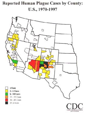
Figure
12. Distribution of plague in the
Western US by County. Image courtesy of
The Center for Disease Control and Prevention.
The symptoms of the disease include a rapid
onset, with an incubation period of only 2 to 10 days, a fever reaching up to
104oF, and flu-like nausea. In some cases, buboes (hence the
name bubonic plague) form from swollen lymph nodes in the pelvic area, groin,
armpits, and back of neck. The buboes
form on the second day after symptoms appear and are very tender and
sore. The disease can then progress to the blood (septicemia plague)
system where the spleen and liver are affected. In the third stage, which
is 90% lethal, the disease progresses to the respiratory tract (pneumonic
plague)? This is an extremely contagious stage and can be transmitted
through coughing. Handling of any wild animals should not be undertaken
unless absolutely necessary. Veterinarians appear to be the group most at
risk for contracting this disease.
Both male and female fleas transmit the disease
while feeding. This occurs due to a plague bacterial buildup in the gut of the
insect and subsequent regurgitation of the blockage by the flea into the host
while feeding. Plague season typically occurs during the summer
months. Apparently higher temperatures
are necessary for a bacterial blockage to occur in the flea’s gut. Because of
the blockage, the flea does not "feel" full and jumps from host to
host in order to feed more. This, of course, spreads the disease even
further. There is a vaccine available, but it is not recommended because
of severe side effects. Currently, only military personnel are given this
vaccine.
Murine Typhus (Rickettsia typhi) is another disease that
fleas transmit and is usually found in rural areas. The fleas responsible
for this disease are the same as those that vector plague. Antibiotics
are effective when given at the first onset of symptoms.
With very heavy prolonged flea infestations pets
can develop anemia. In extreme cases a
pet may harbor an infestation of several hundred fleas. In such cases the animal’s immune system can
be severely damaged resulting in death.
Heavy infestations are more common in kittens and puppies as they are
less proficient than adults in mechanically removing fleas by biting and
scratching.
Pets may develop allergic reactions to flea
bites. This is especially true in older
pets, which have a long history of flea infestations. As with most allergic reactions the tendency
to do so is genetically linked. It is
not uncommon in households with many cats for a few to develop this type of
allergies while others harboring approximately the same number of fleas are
apparently unaffected. In cases of
extreme allergic reactions the animal will loose large amounts of hair,
especially around the rump and on the tail.
One of our black cats (we call him old bald butt Bart) develops this
condition every flea season. Once such
an allergy develops the bite of a single flea can initiate the condition. In addition to hair lost the animal may
develop reddened skin and small scabs over much of his or her body. The latter are partially due to the excessive
scratching of the animal. Normally a
veterinarian can be corrected allergic reactions by injection and oral
administration of cortisones and flea control.
Fleas can serves as an intermediate host
for dog and cat tapeworms. Fleas are
typically infested with the cyst when the larvae feed on the feces of a
tapeworm infested host. The cyst form is
subsequently passed on to the adult flea.
Consuming the adult flea can in turn infest a cat, dog or even small
child. Once consumed the worm form
emerges from the cyst and begins to feed in the digestive tract. Tapeworm feed by absorbing nutrients. This may result in loss of weight and a
rundown condition in the host. An
infestation is usually diagnosed by the presence of the crawling almost square
worms that are about the size of a grain of rice in the feces of the host.
COMMON
FLEAS
The
Oriental rat flea, Xenopsylla cheopis (Figure 13), and northern rat
flea, Nosopsyllus fasciatus are the primary vectors of the
plague. These fleas attack the Norway rat, roof rat, and black rat, all
of which are commonly associated with humans. They also are typically
found on ground squirrels, wood rats, and prairie dogs.
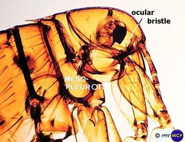
Figure 13.
The Oriental rat flea. Image courtesy of Department of Parasitology,
University of Sao Paulo, Brazil.
The 2 major fleas that are of
household concern are the dog flea, Ctenocephalides
canis, and the cat flea C.
felis, respectively (Figure 2). Both of these are problematic for
pets in the US, with some animals having severe allergic reactions.
Typical symptoms of an allergic reaction to fleas in cats and dogs include a
loss of hair, especially around the upper rump, flaky dry skin and many bumps
and scratches due to the pet’s attempts to remove the fleas. Older animals are typically most
susceptible. This is especially true if
the animal has had a history of flea infestations. In some cases the bite of a single flea can
result in these symptoms if the animal is highly allergic. Treatment for allergic reactions includes
flea control and cortisone shots by a veterinarian.
Typically when these 2 species
bite humans most bites occur around the lower legs (Figure 14). As with some
other fleas when they bite they will feed many more than needed for
nourishment. The bites shown on the
above illustration are those from a few fleas (possibly one).

Figure 13. Typical
cat flea bites occurring on lower legs.
Image Courtesy of Vopak Inc.
The human flea, Pulex irritans (Figure 14), is found all over
the world. Besides humans, it infests
cats, dogs and many other animals, particularly pigs. It breeds profusely in
pigsties, and people working in them can pick up large numbers of fleas and
start infestations in their own homes. The human flea is usually the most
important flea in farm areas; while the bite of cat fleas tends to be
concentrated on the lower part of the legs, those of the human flea may be
spread all over the body.

Figure 14. The human flea. Image
courtesy of Department of Parasitology, University of Sao Paulo, Brazil.
The human flea is the species
that was used in the 1800’s in European flea circuses. They were chosen for their exceptional
ability to jump (over 6 inches high and 13 inches laterally) and supposed
strength. They can pull over 400 time
their own weight. They later fact is
especially impressive since the average human cannot pull much over once their
weight. In the circuses these fleas
performed a variety of acrobatic stunts including pulling tiny carts through
the streets of miniature villages. These
circuses eventually became sideshows in fairs and informal outdoor
markets. These events became known as
flea markets, a term we are all familiar with today.
The
sticktight flea, Echidnophaga
gallinacea, is one of the smaller fleas with males measuring less than 1mm
(Figure 15). The adult flea attaches itself on the head or neck of domestic
fowl, sometimes causing ulcers. Females lay their eggs in the ulcers and the
hatching larvae fall to the ground and feed on decaying plant material.
Sticktight fleas can become quite abundant in poultry yards and adjacent
building. Besides birds they can attack humans, rat, cats, dogs, and many other
mammals. They are potential vectors of plague but their habit of remaining
tightly to one host greatly reduces this possibility.

Figure 15. The
adult of the sticktight flea, a poultry pest.
The chigoe, Tunga penetrans, (Figure
16) is also known as chiggers, jiggers, chique and sand fleas. Some of these latter names can be misleading,
as these are neither true chiggers, which are of course mites nor true sand
fleas, which are actually crustaceans.
This is a tiny burrowing flea that is found in the tropical areas of
North and South America, Africa and the West Indies. It is said to have inspired the sailor’s
saying, “I’ll be jiggered”.

Figure 16. The
adult chigoe, also known as a jigger or sand flea. Image courtesy of Department of Parasitology,
University of Sao Paulo, Brazil.
An infestation of these fleas is
referred to as tungiasis. Gonzalez
Fernandez De Oviedo y Valdes noted the earliest report of tungiasis at the turn
of the 16th Century when Spanish conquerors of the crew of the Santa Maria
were shipwrecked on Haiti and became infested with the disease. A few years
later, the Spanish conqueror Gonzalo Ximenes de Quesada reported an entire
village in Colombia that had been abandoned by its inhabitants due to this
disease. Consequently, his soldiers became so infected with the disease that
they could barely walk. In the 17th Century, Aleixo de Abreu, a Portuguese
physician working in the Brazilian government, provided the world with the
first scientific description.
The
sand flea is normally found in the sandy terrain of warm, dry climates. It
prefers deserts, beaches, stables, stock farms, and the soil and dust close to
farms. While both male and female sand fleas intermittently feed on their
warm-blooded hosts, it is the pregnant female flea that burrows into the skin
of the host and causes the cutaneous lesion. She does not have any specialized
burrowing organs; rather, she simply attaches to the skin by her anchoring
mouth and claws and burrows violently into the epidermis. Since this process is
painless, it is thought that the flea may release some type of tissue
dissolving enzymes. After penetrating she leaves her posterior (rear) end
exposed (Figure 17). The "black
dot" of the nodule is this posterior end of the flea sticking out. The
opening provides the flea with air and an exit route for feces and eggs. With
its head deep into the skin, the flea begins to feed on the host's blood and
enlarges up to 1cm in diameter. Over the next two weeks, over 100 eggs are
released through the exposed opening and fall to the ground. The flea then dies
and is slowly sloughed by the host's skin. The eggs hatch on the ground in 3-4
days. In the next 3-4 weeks, they go through their larval and pupal stages and
become adults. The complete life cycle of these fleas is about one month.
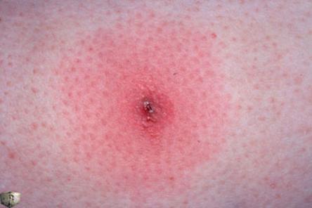
Figure 17. An
adult chigoe buried into the skin.
The first evidence of infestation by this sand flea is a tiny black dot
on the skin at the point of penetration. Because the flea is a poor jumper,
most lesions occur on the feet (Figure 18), often on the soles, the toe webs,
and around or under the toenails. Among natives who frequently squat, however,
the buttocks and scrotal sac can be involved. A small, inflammatory papule with
a central black dot forms early. Within the next few weeks, the papule slowly
enlarges into a white, pea-sized nodule with well-defined borders between
4-10mm in diameter. This lesion can range from asymptomatic to pruritic to
extremely painful. Multiple/severe infestations may result in a cluster of
nodules with a honeycomb appearance. Heavy
infestations may lead to severe inflammation, ulceration, and fibrosis.
Lymphangitis, gangrene, sepsis, the loss of toenails, autoamputation of the
toes, and death may also occur. In most cases, however, this lesion heals
without further complications. Nonetheless, the risk of secondary infection is
high. Tetanus is a common secondary infection that has reported associations
with death. People should avoid walking
with bare feet where these fleas are known to occur. Of course dogs and other pets are frequently
infected (Figure 19).

Figure 18. Ulcers on the feet due to burrowing chigoe
fleas. Image courtesy of Department of Parasitology, Sao Paulo, Brazil.
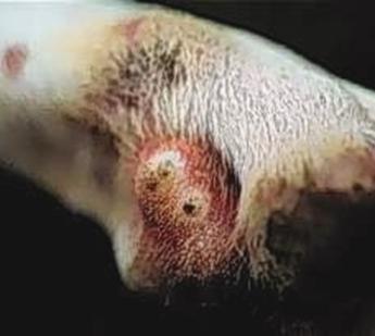
Figure 19. Ulcer
on dog legs due to feeding of chigoe flea.
Image courtesy of Department of Parasitology, Sao Paulo Brazil.
FLEA CONTROL
The cat flea
is the most common flea found feeding on hosts such as domestic pets, humans,
rodents, raccoons, and other wild animals. Successful flea control begins with
identification of the species involved, determining the source of infestation
and understanding the flea life cycle.
The first step is to interviews the building occupants concerning household
practices and recent animal visitors.
This will alert the professional as to hot spots and potential sources
of the infestation. Next a thorough interior inspection should be made to
determine the presence of fleas, concentrating on where domestic animals sleep
or common avenues of travel. This can be accomplished by using a vacuum device,
a white-sock test or a light trap, as well as inspecting domestic animals
thoroughly. Finally a thorough exterior
inspection should be performed, especially where domestic animals frequent or
wild animals nest, such as under porches, crawl spaces, perimeter fence areas,
kennel areas, and overgrown vegetation (shrubbery).
Successful control
typically encompasses eradication of the fleas on the pet, in the home and in
outdoor locations. Control on the pet
alone is typically futile since the eggs, larvae and pupae are found off the
host thus continually providing a source of new adult fleas for reinfestation.
Treatment of Pets.
There are an
infinite number of products and devices available for control of fleas on
pets. Although the pest control operator
typically does not (and should not) become directly involved in this aspect of
flea control, it is to his or her advantage to be aware of the various means
that are available for this purpose.
Flea combs.
These devices are not very effective and when used properly will only
remove 10 to 60% of the adult fleas. This method is also very time consuming.
Shampoos. These products tend to be a temporary
solution to an on-going problem. One
advantage is that they do not only kill adult fleas but also remove dried skin
and fleas feces that eventually fall to the ground and serve as food for the
flea larvae. Typically with flea
shampoos the animal should be thoroughly lathered and rinsed after 15
minutes. There are a variety of products
available containing different active ingredients. Pyrethrins are derived from Chrysanthemum flower
heads. They kill adult fleas quickly but
have essentially no residual activity or lasting effects. Pyrethroids are synthetically produced and
have a longer residual activity.
Carbaryl is a carbamate insecticide that has a long history in pest
control. This material is somewhat toxic
to cats so label instructions should be carefully followed. Citrus peel derivatives, such as D-limonene
are used in some shampoo products, which are fairly mild making them useful for
use on kittens and puppies as well as in household with new babies. However, in some cases cats may exhibit
allergic reactions to these materials.
Pennyroyal oil is another natural product used in pet shampoos. Pulegone, the active ingredient in this oil
can be toxic to animals if misused resulting in any of a number of side effect
including death due to liver failure. If
used label directions should be followed carefully.
Topical
Applications. Some of the newest
products for flea control are imidactloprid (Advantage), fipronil (Frontline)
and (Program). Advantage and Frontline
are spot-on oils and are applies monthly as a small drop to the back of the
neck to prevent removal by grooming. The
products quickly spread over the body due to movement of the animal. They are nontoxic to mammals and kill most of
the fleas on the animal within 24 hours.
Advantage provides approximately 98% control for the first 3 week on
dogs and cats. Control drops slightly
during the 4 week. Frontline provides a
somewhat longer residual activity with a 98% kill for up to 8 and 6 weeks on
dogs and cats, respectively.
Lufenuron (Program) attacks the
problem from a different perspective than Advantage or Frontline. The active ingredient in this product is a
chitin inhibitor. Chitin is the main
protein in the insect’s exoskeleton that gives it strength. As with the other above-mentioned products
Program is usually applied on a monthly basis.
It does not kill the adult fleas but once the fleas feed on the treated
pet the active ingredient prevents chitin from forming in the flea’s eggs. As a result when the eggs are produced they
break due to the lack of chitin in the shell.
In order to be effective Program must be used immediately prior and
during the entire flea season. The idea
is to prevent the flea infestations from developing in the first place.
Flea
collars. Most studies indicate
that flea collars are relative ineffective when compared to many other
available means of control. This is
especially true when ultrasonic flea collars are concerned. These devices have been found to have no
significant effect on fleas.
Control in the Home and Yard
It is worthwhile to discuss with
family members the areas in and around the home where pets spend most of their
time sleeping and resting. Hot spots of
activity can be determined by placing white socks over shoes and walking
throughout the residence. Hot spots in
homes with dogs are usually areas where the animals go in and out of the home,
eat, sleep and spend time with the family.
Cats frequent similar areas but should include high areas including tops
of refrigerators, cabinets and bookcases.
There are commercially available flea traps (Happy Jack and pulvex),
which can be useful for monitoring flea populations. Research indicates that the larva do not move
far from the site of hatching, especially if food is available.
Sanitation
Sanitation is one of the first steps to
effective flea management in the home.
Vacuuming on a regular basis (a minimum of every other day) will remove
many of the various stages, dried blood and other sources of larval food from
the carpet or areas where the pets sleep.
Prior to any pesticide application the carpet should be vacuumed
thoroughly with a beater-bar type vacuum.
This will open up the carpets nap to allow treatment to reach deeper
into the carpet where much of the infestation occurs. Research indicates that approximately 83% of
the larvae spend most of their time deep in the carpet at the base of the
fiber. At pupation the larvae move up
the carpet fiber spinning a camouflaging cocoon around itself. Vacuuming is especially important where lint
and pet hairs accumulate along baseboards, around carpet edges, on ventilators,
around floor heaters, in floor cracks and under and in furniture where pets
sleep. After vacuuming, the vacuum bags
should be discarded in an outdoor trash container. Thorough vacuuming can be an effective tool in a flea pest
management system. All pet bedding
should be thoroughly washed on a regular basis.
Trimming lawns and weeds will create a drier, less-ideal environment for
flea larvae. It can also be beneficial
to avoid piles of sand and gravel around the home. Dogs should be prevented from roaming freely
in and outside the yard, especially in heavily weeded areas or other areas
where they may encounter reinfesting fleas.
Areas that are difficult to access such as crawl spaces or attics under
a home should be sealed to prevent pet entry.
A crawl space is an ideal location for fleas to breed. It is also important to eliminate possible
infestations of wild animals such as raccoons, opossum rats and birds near the
premises.
Chemical Control
Even though much of the
above can useful in preventing and reducing flea infestations in the home
chemical control is the ultimate weapon.
There are dozens of chemicals registered for flea control and a
discussion of all these are beyond the scope of this manuscript. We have tried to include those that are most
commonly used and are not in the business of recommending any.
Before treating, the
homeowner should remove all toys and pillows off the floor or carpet, from
under beds and furniture and on closet floors.
All areas frequented by pets (e.g. table tops, refrigerator tops, window
sills, counter, etc.) should be thoroughly cleaned. All pets should be removed from the premises
prior to treatment. This would include
covering aquariums.
Insect Growth
Regulators (IGRs). This is a relatively new group of chemicals
with a totally different mode of action than the so called conventional
insecticides. Although the growth
regulators do eventually kill their primary function is to disrupt the growth
and molting process. In most cases this results in malformed insects that
cannot function properly. In order to
understand how these products work a brief description of the function of the
hormones that control growth and molting in insects is needed.
Hormones are chemical
substances that are introduced into the blood and subsequently to other parts
of the body where they produce some effect on physiological processes. In insects there are 3 glands that secrete
hormones that regulate growth and molting.
These are the brain hormone, ecdysone and the juvenile hormone
The brain hormone, which is
secreted by a gland at the base of the brain, plays an important role in
molting by stimulating a pair of glands in the prothorax to produce a second
hormone called ecdysone or the molting hormone.
Ecdysone functions to control when molting occurs and the rate of growth
of insects. The third hormone, juvenile
hormone or JH is secreted by another gland in the brain and functions to
determine what an insect will molt into.
If there is a high level of juvenile hormone during the molting process
the insect will stay in a juvenile stage-e.g.-larva to larva, nymph to
nymph. If there is a low level the
insect will advance in stage.
Scientists have chemically
identified some of these hormones in key insects. Once the chemical structure of a hormone is
known it can be synthetically produced in the laboratory. It turns out that if abnormal amounts of a
synthetically produced hormone are applied to an insect then abnormal molts
occur. In some cases monstrous, deformed
insects occur (e.g.-half larva half pupa).
There hasn’t been much done with the brain hormone. It typically has too complex of a molecule,
which makes it too hard to identify or synthetically produce in the laboratory. Ecdysone typically has a steroid component to
its molecule. Since humans also have
steroids in their bodies this might makes it difficult to get these types of
chemicals registered as a useable pesticide with the EPA.
The juvenile hormone is the
main chemical that has been used as an insecticide. Actually the juvenile hormone mimics that are
used do not have the same chemical structure as what occurs naturally in the
insect’s body. The main reason is that a
natural occurring chemical cannot be patented.
As a consequence the chemical companies that produce these pesticides
change the chemical structure a little when they synthetically produced them in
order to patent the compound and protect their investments. As a group the juvenile hormone mimics are
referred to as Insect Growth Regulators.
There are currently a few of
these chemicals that are registered for flea control. One of the disadvantages of some of the IGRs
(for outdoor use) is that they tend to breakdown when exposed to sunlight. However, a recent study using pyriproxfen
(Nylar) reported it to be stable in sunlight when used outdoors for three
weeks. In this study it gave 90% control
of developing fleas. Methoprene (Precor)
is reportedly another effective IGR for flea control.
A disadvantage of these
types of chemical is that they do not kill adult insects which of course do not
molt. As a consequence an adulticide is
also mixed in the spray tank when using IGRs.
Other Chemicals.
One
of the more commonly used and effective indoor flea sprays is a combination of
Precor and Catalyst or Safrotin. Also a
coarse spray (40psi) of diazinon (Knox Out 2 FM), resmethrin (Vectrin), applied
to cracks and crevices of floors, molding and baseboards are said to give good
results. Other flea killers include
tetramethrin (Bio Flea Halt), amorphous silica gel (Drione, Tri-Die),
bendiocarb, (Ficam) and diatomaceous earth (Answer, Organic Plus). Chemicals more commonly used by pest control
operators include Ficam +, Cynoff, Tempo, Delta Guard and Suspend.
Water-soluble sprays are generally used for treating all carpeting and
upholstered furniture. The operator
should always read and follow the label directions closely. If followed correctly potential problems can
be greatly eliminated.
If the pets spend any time outdoor treatment of the yard is needed.
When doing so the preferred breeding locations, areas where the animals spend
most of their time and biology of the fleas should be considered. Again outdoors fleas typically breed in
shaded, moist but not wet locations.
Keeping this in mind it does little good and is a waste of time and
money to treat the whole yard. Cats
using sand boxes and dogs sleeping under shrubs or in crawls spaces or garages
provide a reservoir for fleas. Animal
pens, kennels, doghouses, sandy soil and gravel driveway are other important
sources to consider. Clean and sweep
porches, mow lawns and soak dry soil with water before treating to bring flea
larvae up to the surface. Commonly used
insecticides for outdoors treatment include fenvalerate, carbaryl, diazinon,
popoxur, resmethrin, Knox Out and bendiocarb